PRP and Stem Cells for Horse Tendon Injuries: What a New Controlled Trial Found
A randomized, double-blind, placebo-controlled trial tested a sequential treatment strategy — PRP first, then autologous muscle-derived stem cells — for acute tendon and ligament injuries in sport horses. The results offer some of the strongest clinical evidence yet for this approach.
Tendon injuries are among the most frustrating diagnoses in equine sport. They heal slowly, incompletely, and with a recurrence rate that often exceeds 50% — not because the body fails to respond, but because it responds imperfectly, laying down scar tissue that lacks the mechanical resilience of healthy tendon. For competitive horses, that gap between repair and regeneration can end careers.
Regenerative therapies have promised a better path. Platelet-rich plasma (PRP) has been widely adopted for its immediate availability, but the evidence for PRP alone has been inconsistent — benefits tend to be modest and transient. Mesenchymal stem cells (MSCs) have shown more durable promise, but autologous versions require weeks of cell culture, creating a practical dilemma: treat immediately with something imperfect, or wait for something better?
This study, published in Animals in March 2026, tested a third option: do both, in sequence.
Overview
Researchers at the University of Liège conducted a multicenter, prospective, randomized, double-blind, placebo-controlled trial in sport horses with naturally occurring acute tendon and ligament injuries. Thirty horses completed the study, drawn from five veterinary centers, with lesions of the superficial digital flexor tendon (SDFT), deep digital flexor tendon (DDFT), or suspensory ligament branches confirmed by ultrasound and less than 21 days old at inclusion.
The protocol had two phases. At inclusion (T0), every horse received an intralesional injection of autologous leukocyte-reduced PRP, and a small muscle biopsy was taken from the triceps brachii for cell culture. Over the following six to eight weeks, muscle-derived mesenchymal stem cells (mdMSCs) were expanded under controlled laboratory conditions.
Horses that did not show sufficient clinical improvement after PRP — defined as less than a three-point reduction in total clinical score — were then randomized at week eight (T1) to receive either intralesional autologous mdMSCs (n = 17) or placebo (n = 6). A second injection was administered at week twelve (T2) at the investigator's discretion in most horses. Final evaluation occurred at week sixteen (T3).
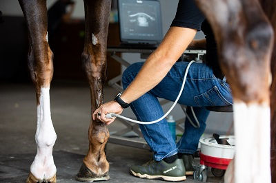
Clinical assessment used a composite total clinical score (TCS) combining lameness grade, pain on palpation, swelling, and a wedge test. Ultrasonographic evaluation tracked fiber alignment, echogenicity, and lesion size as a percentage of tendon cross-sectional area.
Key findings: Evidence for PRP and stem cells for horse tendon injuries
PRP helped — but not reliably enough on its own. Of the 30 horses, seven showed sufficient clinical improvement after PRP alone and were excluded from the randomized phase. That left 23 horses — 77% of the cohort — who did not respond adequately to PRP as a standalone treatment. This aligns with the broader literature suggesting PRP is a useful biological primer but not a consistently disease-modifying therapy for tendon injuries.
Autologous stem cells produced measurable improvement where PRP had not. In the randomized phase comparing mdMSCs to placebo, horses in the treatment group showed significant improvement across clinical and ultrasonographic parameters between T1 and T2. Total clinical score dropped from 6.0 to 4.0 in the mdMSC group (p = 0.0005), while the placebo group remained essentially unchanged (6.0 to 5.0, not significant). Between-group comparison confirmed the superiority of mdMSCs for TCS (p = 0.017). Lameness grade, wedge test response, fiber alignment, and lesion size all improved significantly in the treatment group.
A second injection provided additional benefit. Among the 18 partial responders who received a second mdMSC injection at T2, further significant improvements were recorded by T3 across multiple parameters: total clinical score, lameness grade, wedge test, fiber alignment score, and lesion size all continued to improve. This suggests tendon regeneration with mdMSC therapy is progressive and cumulative rather than a single-dose effect.

The treatment was safe. No systemic adverse events were recorded. Local reactions — mild swelling or discomfort at the injection site — were transient and resolved without intervention. The autologous approach avoids the immune recognition problems increasingly documented with allogeneic stem cell products, where mismatched donor cells can trigger antibody responses and complement-mediated destruction.
The sequential strategy resolved the timing dilemma. By treating immediately with PRP and simultaneously collecting muscle tissue for cell culture, the protocol ensured horses received early biological support while autologous cells were being prepared. The delay inherent in autologous cell therapy — previously a barrier to clinical adoption — was converted into a feature rather than a problem.
How to apply these findings
Understand what PRP can and cannot do. PRP is widely used and readily available, and it appears to provide genuine early biological support — reducing inflammation and priming the lesion environment. But this study adds to a body of evidence suggesting it is not sufficient on its own for many horses with acute tendon injuries. If your vet recommends PRP for a tendon injury, it is a reasonable first step; just understand it may not be the last one needed.
Ask about the full regenerative pathway, not just the first injection. If your horse sustains an acute tendon or ligament injury, the conversation with your vet should include what happens if PRP doesn't produce adequate improvement. Knowing that autologous stem cell therapy exists — and that the cell collection can happen at the same time as the PRP injection — is worth understanding before an injury occurs, not after.
Recurrence risk is the real enemy. With reinjury rates historically above 50% for horses managed conventionally, the goal of regenerative therapy is not just return to work — it is return to work with structurally sound tissue. Ultrasonographic follow-up is not optional; it is how you know whether healing is progressing as it should. A horse that looks sound may still have a lesion that is not ready for full work.
Preventive cell banking is an emerging option. The researchers note that the minimally invasive microbiopsy technique used in this study opens the possibility of banking autologous stem cells in healthy, high-performance horses before any injury occurs. If your horse competes at a high level, this may be worth discussing with a veterinary sports medicine specialist — having cells already prepared removes the six-to-eight-week production delay entirely.
Rehabilitation still matters. Even in this study, the researchers acknowledged that rehabilitation protocols varied because these were client-owned horses in real clinical settings. The biology of tendon healing can be supported by stem cells, but it cannot be replaced by them. Controlled exercise progression, monitoring, and patience remain foundational to successful outcomes.
Read the study
Serteyn, D., Graide, H., Ceusters, J., Vandersmissen, M., Salciccia, A., Sandersen, C., & Lejeune, J.-P. (2026). Sequential application of autologous platelet rich plasma and muscle-derived mesenchymal stem cells for acute tendon injuries in horses: Early clinical and ultrasonographic outcomes in a randomized, double-blind controlled study. Animals, 16(6), 940. https://doi.org/10.3390/ani16060940


Comments